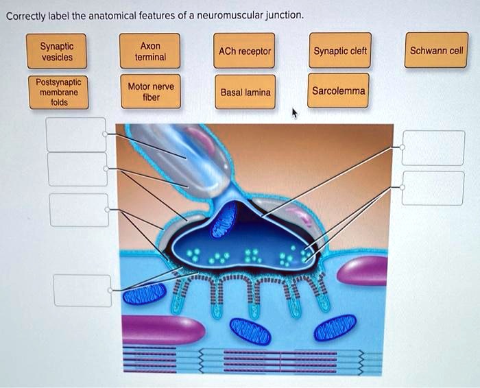
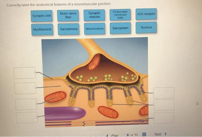
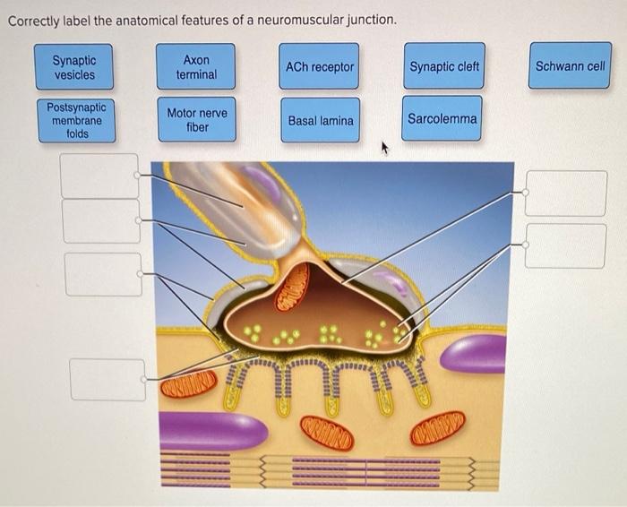
Correctly label the anatomical features of a neuromuscular junction. – Correctly labeling the anatomical features of a neuromuscular junction (NMJ) is essential for understanding its structure and function. This guide provides a comprehensive overview of the NMJ, its components, and the methods used to label them accurately.
The NMJ is a specialized synapse that transmits nerve impulses to muscles. It consists of a presynaptic terminal, synaptic cleft, postsynaptic membrane, and motor end plate. Each component plays a crucial role in the process of neurotransmission.
Introduction to Neuromuscular Junction
The neuromuscular junction (NMJ) is a specialized synapse that transmits nerve impulses from motor neurons to skeletal muscle fibers, enabling voluntary movement. It comprises a presynaptic terminal, synaptic cleft, and postsynaptic membrane.
Anatomical Features of NMJ

Presynaptic Terminal:The presynaptic terminal contains synaptic vesicles filled with neurotransmitters (acetylcholine) that are released upon nerve impulse arrival. Synaptic Cleft:The synaptic cleft is a narrow gap separating the presynaptic and postsynaptic membranes. Calcium ions entering the presynaptic terminal trigger neurotransmitter release into the synaptic cleft.
Postsynaptic Membrane:The postsynaptic membrane contains acetylcholine receptors that bind to acetylcholine released from the presynaptic terminal, triggering muscle contraction. Motor End Plate:The motor end plate is a specialized region of the postsynaptic membrane where acetylcholine receptors are concentrated. Muscle contraction is initiated at the motor end plate.
Importance of Correct Labeling
Accurate labeling of NMJ components is crucial for understanding its structure and function. Mislabeling can lead to errors in research and clinical practice, potentially affecting the diagnosis and treatment of neuromuscular disorders.
Methods for Labeling NMJ

Immunohistochemistry:Immunohistochemistry uses specific antibodies to label NMJ components. This technique allows for precise localization and visualization of proteins within the NMJ. Electron Microscopy:Electron microscopy provides detailed ultrastructural images of the NMJ, revealing its subcellular organization and molecular composition. Electrophysiology:Electrophysiology records electrical activity at the NMJ, allowing for the study of neurotransmitter release, receptor function, and muscle contraction.
Applications of NMJ Labeling

Research:NMJ labeling has aided in understanding neuromuscular diseases, such as myasthenia gravis and amyotrophic lateral sclerosis. It enables the identification of molecular defects and the development of therapeutic strategies. Clinical Practice:NMJ labeling assists in diagnosing and treating neuromuscular disorders. Muscle biopsies can be labeled to identify abnormalities in NMJ structure and function, guiding clinical decisions.
FAQ: Correctly Label The Anatomical Features Of A Neuromuscular Junction.
What is the role of the presynaptic terminal in the NMJ?
The presynaptic terminal releases neurotransmitters into the synaptic cleft, which triggers muscle contraction.
What is the function of the synaptic cleft?
The synaptic cleft is the space between the presynaptic and postsynaptic membranes, where neurotransmitters are released and bind to receptors.
What is the role of the postsynaptic membrane in the NMJ?
The postsynaptic membrane contains receptors that bind to neurotransmitters, triggering muscle contraction.